What are the types of Diabetic Retinopathy?
There are 2 main stages of diabetic retinopathy:

Non-Proliferative Diabetic Retinopathy (NPDR)
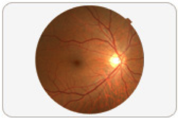
NPDR is an early stage of the disease where the blood vessels in the eye become leaky. At this stage, vision is usually preserved. The blood vessels can also leak fluid or proteins, resulting in swelling of the retina. If the swelling occurs at the centre of the retina (also known as the macula), this is called maculopathy. In these situation, central vision can be affected.

Proliferative Diabetic Retinopathy (PDR)
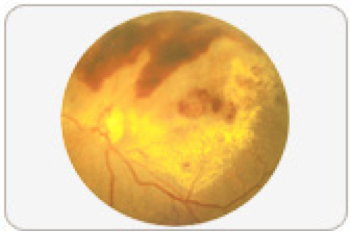
PDR is a late stage of the disease where new but abnormal blood vessels are formed. These fragile vessels bleed easily and result in a leakage of blood in the eye, known as vitreous haemorrhage. With time, these vessels scar and pull the retina off the eye. This is known as tractional retinal detachment and may lead to blindness. If the blood vessels grow on the iris, this can lead to increased pressure in the eye known as glaucoma. This will cause pain and eventual damage to the nerve of the eye and blindness.
Symptoms and Diagnosis
In the early stages of NPDR (Non-Proliferative Diabetic Retinopathy), vision is usually not affected. At this stage, the retinopathy is often not diagnosed unless the patient goes for regular screening. When the macula gets affected, (a condition known as maculopathy), the patient will experience some blurring and/or distortion of vision.
In the late stages also known as PDR (Proliferative Diabetic Retinopathy), the patient may still not experience any symptoms. The patient will experience sudden onset of blurring or loss of vision or floaters when vitreous haemorrhage occurs. When the condition progresses on to retinal detachment this may be associated with an increase in floaters, flashes of light or darkening of a certain portion of the vision.
To find out more about Diabetic Retinopathy, book an appointment with us
At EEC, we can perform a complete ocular examination including visual acuity, slit lamp, intraocular eye pressure checks and dilated fundal examination.
With the latest imaging techniques including FFA, we offer an accurate diagnosis and staging of diabetic retinopathy and a state-of-the-art spectral domain OCT, Zeiss Cirrus and subsequiently personalized treatment for your condition after your assessment.
